Module 1 — Neuroscience 101

What is neuroscience? How do MRI and fMRI map the brain — and how can tDCS/TMS safely nudge networks?

Concept

Structural MRI

Read anatomy like a map: slices, landmarks, and lingo.

Concept

Functional MRI

Activity overlays (BOLD) — what “activation” really means.

Tool

tDCS

Gentle current via scalp electrodes to modulate networks.

Tool

TMS

Brief magnetic pulses that activate cortex — “the click.”

Practice

Vocabulary

Cortex, voxel, BOLD signal, montage, target.

Activity

Mini-task

Landmarks, reading an activation map, and targeting ideas.


What is “Neuroscience,” really?

Neuroscience is the study of the nervous system — how the brain is built (structure) and how it works (function).

In this program, you’ll use:

  • Structural MRI → high-resolution pictures of anatomy (the map).
  • Functional MRI (fMRI) → color overlays showing where activity increases during tasks (the traffic).
  • tDCS → gentle, non-invasive currents via scalp electrodes to modulate networks.
  • TMS → magnetic pulses that briefly activate cortex in a precise spot (“the click”).
Myth-buster: fMRI is not mind reading; it measures oxygen-level changes (BOLD) related to neural activity.
Want to know more about neuroscience? (books & video)

Foundational Neuroscience Textbooks

  • Principles of Neural Science — Kandel, Schwartz, Jessell.
  • Neuroscience: Exploring the Brain — Bear, Connors, Paradiso.
  • Fundamental Neuroscience — Squire et al.
  • Neuroanatomy through Clinical Cases — Blumenfeld.

Video resource

Fundamentals of Cognitive Neuroscience — Dr. Hamed Ekhtiari
👉 LINK

Why these tools together?
  • Maps + effects: MRI/fMRI tell you where/when; tDCS/TMS test what happens if we nudge here.
  • Non-invasive & safe: we follow screening, intensity, and duration guidelines. No surgery, no shocks.
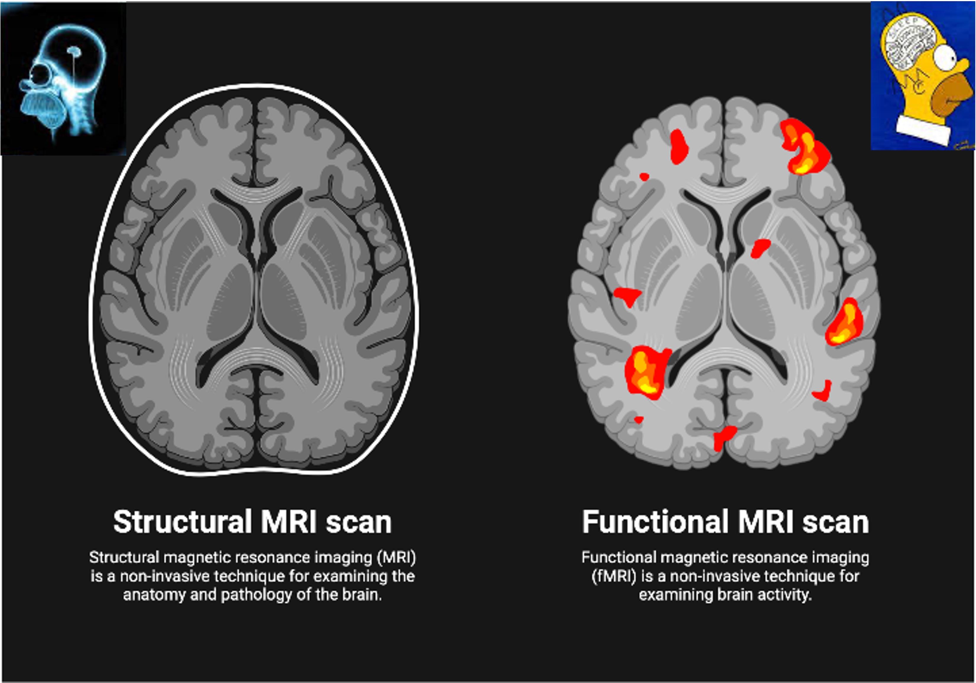
Quick look: one brain, two views (figure)
Structural MRI with fMRI overlay

Structural MRI = the map. fMRI = activity patterns during a task/rest.

Mini-activity (10–15 min)
  1. Spot the landmarks. Find frontal, parietal, temporal, occipital lobes on structural MRI.
  2. Read an activation map. Identify a “hot” area on a simple fMRI overlay.
  3. tDCS thought experiment. If your goal is attention, which lobe might you consider? Why?
  4. TMS target idea. Where would you place the crosshair for a motor task demo?
Write 3–5 sentences answering 2–4. Save them for your Module 1 deliverable.
Vocabulary (click to expand each term)
Cortex

Outer folded layer of the brain where many higher functions occur.

Voxel

A 3D pixel in MRI images (volume element).

BOLD signal

fMRI’s oxygen-linked activity measure (Blood-Oxygen-Level Dependent).

Montage

tDCS electrode arrangement (positions + current).

Target

The specific cortical spot selected for TMS.

What you’ll turn in (Module 1)

Create a Markdown file in your fork: docs/work/module1/<your-username>-notes.md with:

  • 3–5 sentences from the mini-activity.
  • One vocabulary term explained in your own words.
  • One question you’re curious about next.

Commit on branch m1-<your-username> and open a PR titled [M1] Notes and reflection — <your-username>.


← Back: Module 0 🏠 Main Page → Next: Module 2